Are you wondering who discovered microscope? Let’s dive into this fascinating topic and uncover the story behind this groundbreaking invention! The microscope is an essential tool that has revolutionized our understanding of the world by magnifying tiny objects and revealing intricate details invisible to the naked eye. But who discovered microscope and made this incredible leap in science possible? In this article, we’ll explore not only who discovered microscope but also how it works, its evolution over time, and its profound impact on scientific discovery.
The journey to understanding who discovered microscope takes us back to the 16th and 17th centuries when pioneers like Hans and Zacharias Janssen and later Antonie van Leeuwenhoek transformed the way we see the world. Imagine how life-changing it must have been to view tiny organisms for the first time! If you’re still curious about who discovered microscope and how their invention changed the course of history, you’re in for a treat. By the end of this article, you’ll have a clear understanding of not just who discovered microscope but also why it remains one of the most significant inventions in science and medicine to this day!.
A microscope is a fascinating instrument that magnifies objects, allowing us to see details that are invisible to the naked eye. It has transformed the way we explore the world, especially in science and medicine. Have you ever wondered who discovered microscope? In this article, we’ll dive deep into the history and evolution of this revolutionary tool, uncovering the answer to who discovered microscope and how it has advanced over time. Let’s begin this exciting journey to learn more about microscopes and, most importantly, who discovered microscope!
The microscope, a tool that allows us to peer into the unseen world, has a rich and fascinating history. From its humble beginnings to the powerful instruments used today, here’s a glimpse into the evolution of this groundbreaking invention:
The microscope, a tool that allows us to peer into the unseen world, has a rich and fascinating history. From its humble beginnings to the powerful instruments used today, it’s worth exploring who discovered microscope and how it evolved over time. Have you ever wondered who discovered microscope and made it possible to see the intricate details of life? The journey begins in the late 16th century, with Hans and Zacharias Janssen often credited as the pioneers. As you delve deeper into the story, you’ll learn not only who discovered microscope but also how their invention sparked a revolution in science and medicine. Here’s a glimpse into the evolution of this groundbreaking invention:
The microscope has revolutionized our understanding of the world around us, from the building blocks of life (cells) to the invisible world of microorganisms. Each inventor and their contribution, from the early compound microscopes to the modern DIY versions, played a vital role in this incredible scientific journey.
While Zacharias Janssen is known for the invention, other scientists improved it significantly:
| Scientist | Contribution | Year |
|---|---|---|
| Galileo Galilei | Developed his own version called the “occhiolino” (little eye) | ~1609 |
| Robert Hooke | Improved the microscope and coined the term “cell” | 1665 |
| Anton van Leeuwenhoek | Built powerful single-lens microscopes and discovered microorganisms | 1670s |
The microscope, a groundbreaking scientific instrument, evolved through contributions from various researchers. In 1590, Hans and Zacharias Janssen became the first who discovered microscope, laying the foundation for modern microscopy. Later, in 1670, Antonie van Leeuwenhoek advanced the field by creating the first microscope for scientific research and becoming the first to observe bacteria.
While often confused with microscopes, magnifying glasses are simpler tools that provide low magnification by zooming in on objects. Unlike loupes, which are used close to the eye, magnifying glasses are larger and designed for use at a distance, typically with a focal length of at least 125 mm. Understanding who discovered microscope helps us appreciate the leap from basic magnification tools to the advanced instruments we use today. Thanks to who discovered microscope, our view of the microscopic world has been transformed forever!
Such a gadget generally comprises a single lens that reflects light to change its direction. In general, the lens is fixed inside a frame for stability.
Let’s know its limitations:
| Date | Inventor(s) | Microscope Type | Magnification (approx.) |
|---|---|---|---|
| Late 1500s/Early 1600s | Zacharias Janssen (possibly with Hans Janssen) | Early Compound Microscope (bi-convex eyepiece/plano-convex objective) | Low power (3x to 9x) |
| Late 1600s | Robert Hooke/Christopher Cock | Compound (bi-convex eyepiece and objective with removable field lens) | Up to 50x |
| 1609 | Galileo Galilei | Compound (bi-concave eyepiece/bi-convex objective) | Up to 30x |
| 1676 | Antonie van Leeuwenhoek | Simple (single bi-convex lens) | Very high for the time (up to 270x) |
| Late 1800s/Early-Mid 1900s (widely manufactured) | Various | Dissecting Microscopes (Simple) | Low to moderate power |
| Present Day | DIY Enthusiasts | 2D/3D Printed Simple Microscopes | Low power |
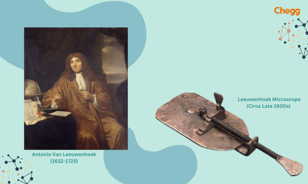
In 1665, Antonie Van Leeuwenhoek, often celebrated as the father of microscopy, created a microscope that allowed him to observe previously unseen particles, including bacteria. But who discovered microscope that paved the way for this groundbreaking work? While Hans and Zacharias Janssen are credited with inventing the early compound microscope, Leeuwenhoek’s innovations elevated its use to new heights.
Antonie Van Leeuwenhoek mastered the art of machining and polishing lenses, creating an advanced lens capable of magnifying up to 270x. Have you ever wondered who discovered microscope and how it enabled Leeuwenhoek to achieve such incredible breakthroughs? Through his work, Leeuwenhoek became the first person to observe live cells, including bacteria, sperm, and red blood cells. He even discovered microorganisms in saliva and rainfall, naming them “animalcules.”
If you’re curious about who discovered microscope and how this invention evolved to change science forever, Leeuwenhoek’s story offers a fascinating chapter in the history of microscopic exploration.
Even though the Leeuwenhoek microscope only had one lens, it had better clarity and magnification than comparable compound microscopes.
They crafted the frames of the Van Leeuwenhoek microscope from copper, bronze, or occasionally silver. They consisted of two plates holding a single lens in line, accompanied by a small hole. A sample was placed on a clip in the lens’s viewing area and fixed onto a block. The sample’s height in its area of vision and distance from the lens were both adjustable with two screws.
Perhaps his most famous experiment came in 1674 when he viewed some lake water:
“I now saw very plainly that these were little eels, or worms, lying all huddled up together and wriggling just as if you saw, with the naked “eye, a whole tubful of little eels and water, with the eels squirming among one another; and the whole water seemed to be alive with these multifarious animalcules.
This was for me, among all the marvels that I have discovered in nature, the most marvelous of all, and I must say, for my part, that no more pleasant sight has ever yet come before my eyes than these many thousand living creatures seen all alive in a little drop of water, moving among one another, each several creatures having its proper motion.”
Antonie van Leeuwenhoek did some amazing things to help make microscopes better. Here’s what he did:
So, even though Leeuwenhoek didn’t invent the microscope, he made it a lot better. His work helped start the study of tiny living things, called microbiology. His love of learning and his hard work helped us understand more about the tiny world around us.
Scientists have developed various microscopes, each becoming more advanced and serving different purposes. Here are the main types:

A microscope is an instrument that includes different components that have their functions. The workings of a microscope depend on its structure. Let’s study the structure of the microscope, which also consists of two parts:
Structural parts are the main parts of a microscope. It helps the microscope to work or move properly. These microscope parts work together to magnify objects that we cannot see with our naked eye. The structure of the microscope includes three parts:
Head: The microscope’s main part contains lenses through which we can see the object.
Arm: It connects the base and head, which are used to carry the microscope.
Base: The base provides support and stability for the microscope.
The optical microscope parts work together to ensure the proper functioning of a microscope. A sample put on a slide is seen, magnified, and imaged using the optical components of the microscope. It has several components:

A compound microscope, a revolutionary tool, uses two lenses to significantly enhance the magnifying power, making it possible to observe minute details of objects. But have you ever wondered who discovered the microscope that laid the foundation for this innovation? This invention has had a profound impact on various fields, including pathology and forensics.
In pathology labs, the compound microscope is used to identify disease-causing bacteria and diagnose illnesses, a practice made possible thanks to who discovery microscope. Similarly, in forensic labs, human tissue and food samples collected from crime scenes and courtrooms are analyzed under a microscope to identify evidence and solve crimes. Who invented the compound microscope?
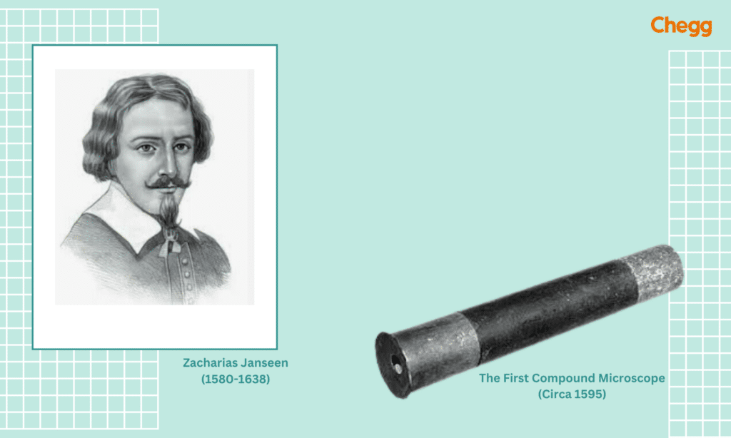
Zacharias Janssen was the first person who discovered the microscope. In 1590, he invented the first-ever compound microscope. Many scholars believe that Hans Janssen made a significant contribution, despite Zacharias Janssen being just a teenager in the 1590s and being credited with developing the compound microscope. Hans Janssen was an eyeglass maker in Middleburg, Holland. People started using eyeglasses more often during that period, which brought a lot of interest to optics and lenses. In reality, a few scholars claim that Janssens and Hans Lippershey, an old Dutch eyeglass manufacturer, created the microscope simultaneously and independently.
They were a combination of two talents: Janssen was good at doing experiments, and Hans was good at making eyeglasses. So, they combined both things and invented the first microscope. They invented a microscope by putting two lenses on the tube; those lenses make the object. The tube became a microscope with those two lenses that zoomed in on the small objects.
The simple microscope, a revolutionary tool that unveiled the microscopic world, was invented by Dutch scientist Antonie van Leeuwenhoek. While many wonder who discovered the microscope, Leeuwenhoek’s work stands out for his groundbreaking discoveries using his unique design.
Unlike modern microscopes, his simple microscope used a tiny glass bead as a lens, capable of magnifying objects up to 270 times. Though Hans and Zacharias Janssen are credited as those who discovered the microscope, Leeuwenhoek advanced its potential with over 500 meticulously crafted designs. Understanding who discovered the microscope highlights how Leeuwenhoek’s innovations transformed science forever.
The invention of the microscope is attributed to Dutch spectacle maker Zacharias Janssen and his father, Hans, who discovered the microscope in the late 16th century. While they weren’t from India, this revolutionary invention paved the way for innovations across the world, including in India. A notable Indian contribution is by Manu Prakash, an alumnus of IIT Kanpur, who invented the “Foldscope,” a microscope costing less than a dollar. This affordable and portable microscope demonstrates how science can be made accessible to everyone, regardless of their economic background.
But have you ever wondered who discovered the microscope and how it led to such innovations? While the Janssens are credited as those who discovered the microscope, India’s advancements, like Manu Prakash’s Foldscope, highlight the continued evolution and accessibility of this essential tool. Isn’t it fascinating how a centuries-old invention continues to inspire groundbreaking developments worldwide?
Do you know the name of the science that Discovered a Microscope & involves using a microscope? Microscopy is the science of using a microscope. The scientific field of microscopy includes using microscopes to observe items and parts of objects that are not visible to the human eye.
The microscope clears the image of a sample, but microscopy is a science in which scientists and researchers come to an outcome by studying the cells or specimens.
Beyond what the human eye can see, it increases magnification, allowing users to explore the microscopic world fully. Coaxial (or shadow-free) lighting techniques enable clear and error-free visualization of crucial anatomy. Current designs and innovative coating processes in lenses achieve a level of realism never seen before. These advanced techniques made microscopy easy.
As time passed, scientists developed the microscope. Modern technologies aid in the creation of advanced microscopes, which incorporate both high-resolution and super-resolution imaging methods in advanced microscopy techniques. The three branches of advanced microscopy are:
The main 3 branches of advanced microscopy are optical, electron, and scanned probe. Let us tell you about each branch in detail.
It is a common method in biology and medicine. By carefully obtaining the light signals from various angles inside the sample and combining the plane pictures to produce three-dimensional images, this method makes it possible to examine biological samples. Studying samples in three dimensions makes it easy for researchers to give an appropriate result.
This method of microscopy, which is the most popular and well-known, includes enlarging the image of the item by shining light through it or reflecting light off of it, and then studying this light through one or more lenses. Recording the image on a photographic plate or examining it directly on a computer screen is now possible.
Electron microscopy is a science in which you can observe more than a million times smaller details. Except for the fact that they “image” the sample and learn about its composition and structure using a focused flow of electrons rather than photons, electron microscopes (EMs) operate similarly to their optical counterparts.
Microscopes, those amazing tools that let us see the tiniest details invisible to the naked eye, have been revolutionary for science. But why exactly are they so important? Here’s a breakdown of their crucial role in scientific exploration:
Modern microscopes go beyond simple magnification. Electron microscopes, for example, can magnify objects millions of times, revealing details far beyond the capabilities of light microscopes. These advancements continue to push the boundaries of scientific exploration.
Microscopes are crucial tools in a wide range of scientific fields, allowing scientists to see things that are invisible to the naked eye. Here’s a glimpse into how microscopes are used across various disciplines:
These are just a few examples of how microscopes are used across various fields. As technology continues to advance, microscopes are becoming even more powerful and sophisticated, opening doors to discoveries and innovations in numerous scientific disciplines.
Imagine trying to study the building blocks of life, like cells, without a microscope. They’re simply too tiny to see! The microscope allows you to:
While Janssen gets credit for the early microscope, another scientist named Antonie van Leeuwenhoek made significant improvements in the 1600s. He’s often called the “Father of Microbiology” for his groundbreaking discoveries using powerful microscopes.
The invention of the microscope marked a pivotal milestone in scientific history, pioneered by Dutch spectacle makers Zacharias Janssen and his father. Hans around 1590, who crafted the first compound microscope using two lenses in a tube. Building upon this foundation, Antonie van Leeuwenhoek elevated microscopy in the 1670s with his meticulously polished single-lens microscopes, enabling the first observations of living cells, including bacteria, protozoa, and blood cells.
Read More:-
Scientists created this instrument to observe specimens more closely. It magnifies objects, aiding in improved research by making them appear larger.
Antonie van Leeuwenhoek is considered the father of the microscope. In the 1670s, he perfected single-lens microscopes with exceptional magnification and used them to observe bacteria, protozoa, blood cells, and other microorganisms for the first time. His groundbreaking work revolutionized microbiology and greatly expanded our understanding of the microscopic world.
There are two types of parts in a microscope:
Structural parts are visible in the body of the microscope.
Optical parts: those parts combine for the proper functioning of the microscope.
1. Scanning electron microscope
2. Fluorescence microscope
3. Confocal microscopy
4. Electron microscope
5. Optical microscope
The first microscope was invented around 1590 by Dutch spectacle makers Zacharias Janssen and his father, Hans Janssen. They created the compound microscope by placing multiple lenses in a tube, which could magnify objects more than simple magnifying glasses. This invention opened the door to exploring the microscopic world and laid the foundation for modern scientific discoveries.
People often credit Antonie van Leeuwenhoek as the first person to use a microscope for scientific purposes. He made significant contributions to the field of microscopy and was the first to observe and experiment with microbes.
No, Galileo Galilei did not invent the microscope. He is best known for his work with telescopes.
Antonie van Leeuwenhoek made the best simple microscopes to see tiny things. Later, Ernst Ruska invented the electron microscope to see even smaller details, and Gerd Binnig and Heinrich Rohrer made the scanning tunneling microscope to look at atoms.
Richard Adolf Zsigmondy discovered the ultra-microscope in 1903. This special microscope helps scientists see tiny particles that regular microscopes can’t show.